


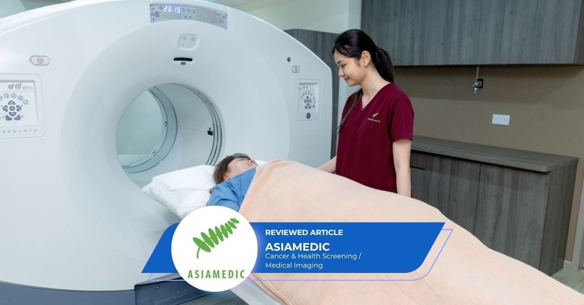
Medical imaging comprises various techniques to view the inside of the body. This allows doctors to find indications of a health condition, diagnose the condition, or to see how well a patient is responding to treatment. Medical imaging tests include X-Ray, Ultrasound, CT scans, MRI scans and PET scans. These are mostly non or minimally invasive. This Health365 Guide to medical imaging explains the different imaging technologies – what they are, the differences, and what they are generally used for.
[Updated Oct 2026]
Contact MedicalTravel.asiaBack to Main Page: Medical Imaging
What Are The Different Medical Imaging Technologies?
Medical imaging technologies that patients commonly come across include x-rays, ultrasounds, CT scan, MRI scans, and PET scans.
What Is An X-Ray?
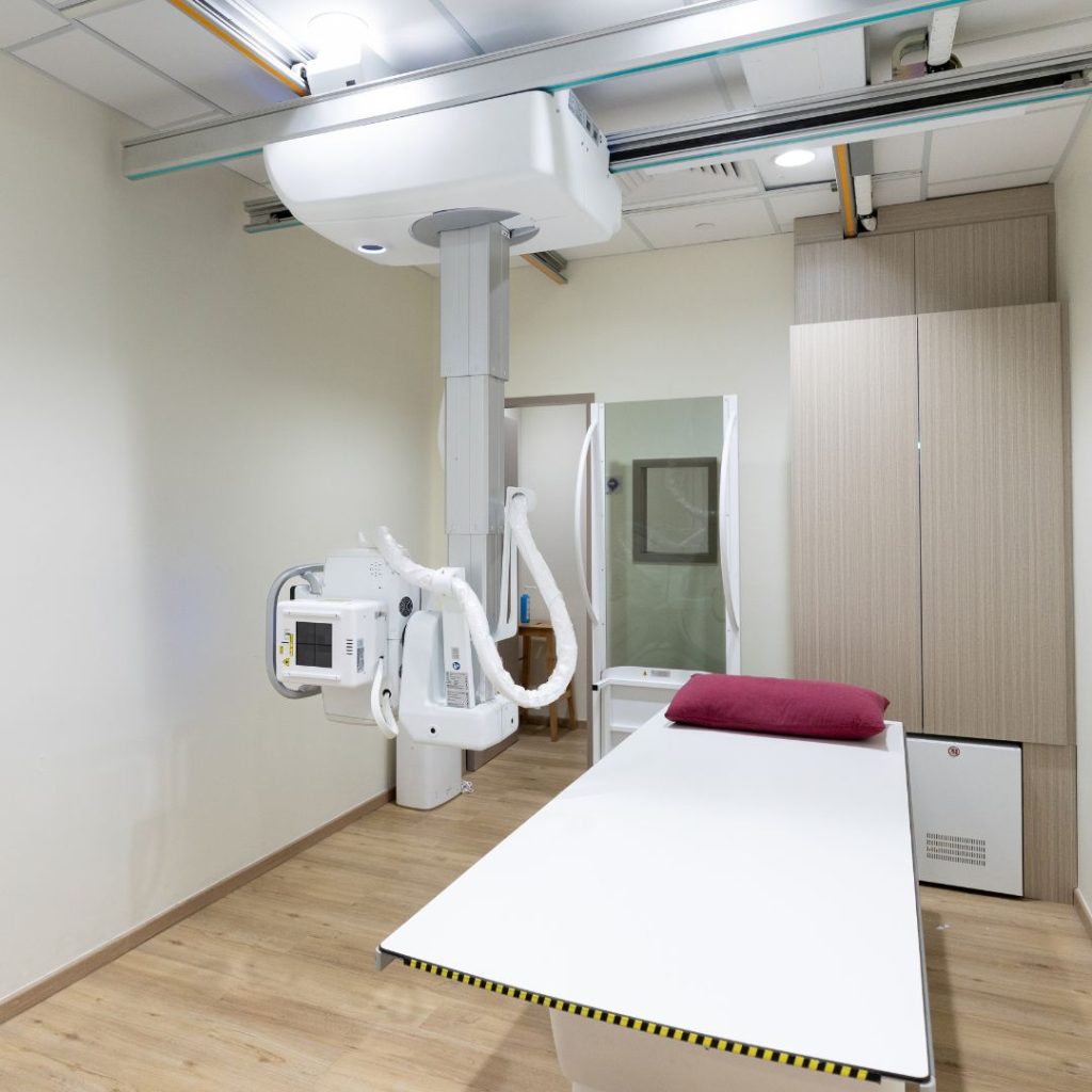
X-ray machines use X-rays, a form of ionising electromagnetic radiation, to produce images of structures inside the body. Standard X-rays may be used for a variety of reasons, including diagnosing tumours or injuries to bones. X-ray scans expose patients to radiation.
- Soft body tissues such as blood, skin, fat, and muscle allow more X-ray to pass through, resulting in darker images on the film or digital media.
- Bones are denser and allow less of the X-rays to pass through, resulting in white images.
- For example, if there is a break in the bone, more of the X-rays pass through the broken area and the break is seen as a dark line against the white bone.
X-rays are also used in other types of diagnostic imaging, such as CT scans.
What Is An Ultrasound?
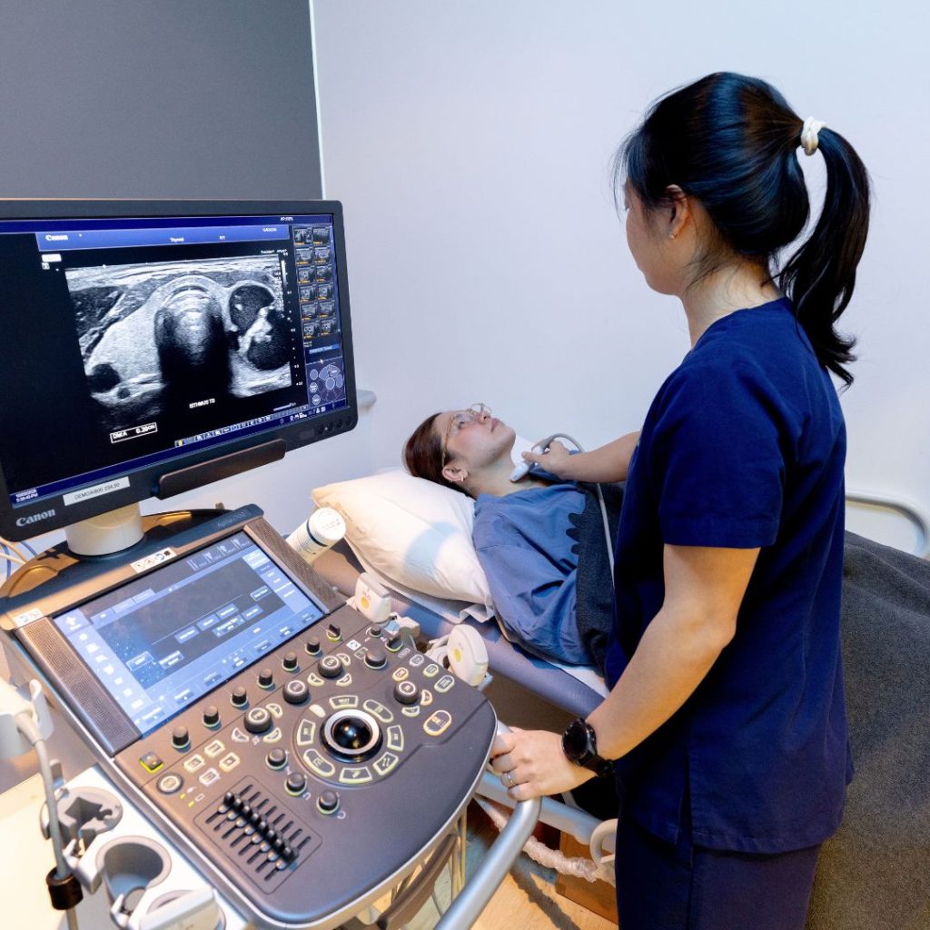
Another diagnostic imaging technology is ultrasound. This non-invasive imaging test uses high-frequency sound waves to create pictures or videos of internal organs or other soft tissues in real-time. Ultrasound does not involve the use of radiation. Ultrasound can help the doctor investigate a variety of conditions, such as unexplained pains, lumps, or the causes of abnormal blood tests.
- During an ultrasound, a transducer is placed over an area of the body. The ultrasound waves are transmitted from the transducer into the body.
- The sound waves bounce off structures inside the body and back to the transducer. A computer then converts the signals into real-time images or videos.
What Is A CT Scan?
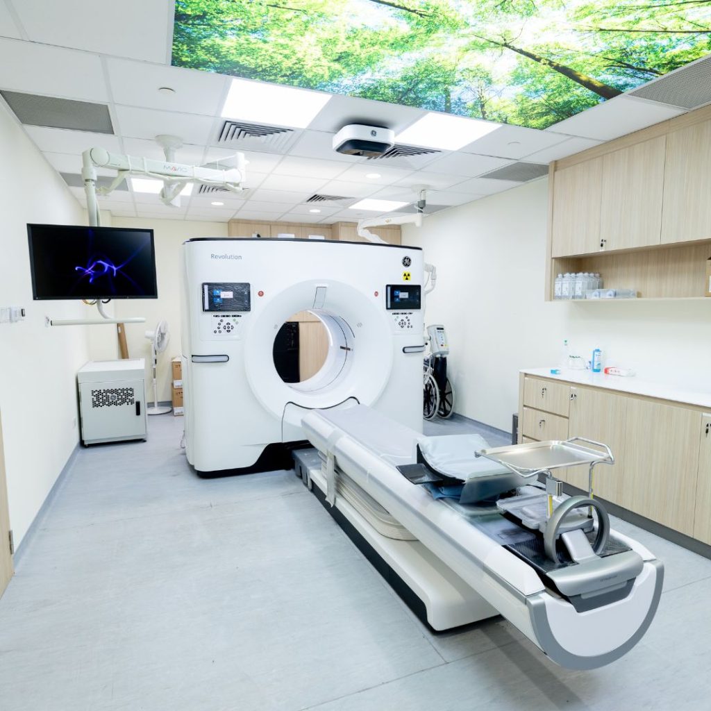
CT (computed tomography) scans use X-rays to produce detailed images of the inside of the body. They are often used to diagnose cancer, heart disease, injuries from trauma, and musculoskeletal disorders. Like standard X-Rays, radiation is used in CT scans.
• Multiple cross-sectional images of the body are taken and re-assembled in a computer to show the internal organs and other structures (e.g. bones, blood vessels).
• Can be rotated to allow the radiologist to view it from various angles.
What Is A MRI Scan?
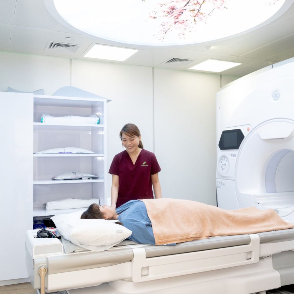
MRI (magnetic resonance imaging) creates detailed images of organs and other structures inside the body using a powerful magnet and radio waves. Patients undergoing this test must remove metal items and inform the technician of any internal devices (e.g. pacemaker) that contain metal. MRI is used to diagnose conditions including ligament injuries and tumours.
• MRI scans may require a contrast agent that is injected through an IV line to allow a better view of the area under study.
• Like CT scan, MRI shows the structure of the body, but produces different set of information.
• Takes longer to perform than a CT and is more expensive.
• Does not expose patients to radiation.
What Is A PET Scan?
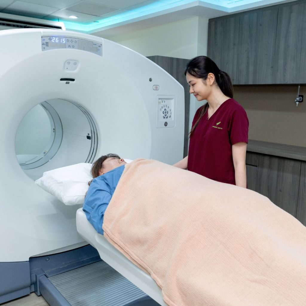
A PET (positron emission tomography) scan is used to visualise the metabolism or chemical activity within the body. Unlike CT and MRI scans that look at the form of structures inside the body, a PET scan looks at the functioning of the body and detected unusual cellular activity. A PET scan may be used to diagnose cancer, heart disease, and brain disorders such as dementia or Alzheimer’s.
- It involves injecting a small amount of radioactive substance (radiotracer) through an IV line into the body.
- The PET scanner detects and records the activity of the radiotracer as it moves through the body.
- PET may detect disease-related changes at the cellular level before they become apparent on some other imaging tests.
PET scans may often be combined with a CT scan for a more complete picture (PET-CT Scan).
| X-Ray | Ultrasound | CT Scan | MRI | PET Scan | |
| What Does It Show? | Structure of tissues / organs / bones. | Structure of tissues / organs / bones. | Structure of tissues / organs / bones. | Structure of tissues / organs / bones. | Metabolism or chemical activity in the body. |
| What Are The Common Diagnostic Uses? | Tumours or injuries to bones. | Unexplained pains, lumps, or the causes of abnormal blood tests. | Cancer, heart disease, injuries from trauma, and musculoskeletal disorders. | Ligament injuries, brain disorders, and tumours. | Cancer, heart disease, and brain disorders. |
| Radiation Exposure? | Yes. | No ionising radiation. Uses sound waves. | Yes. | No ionising radiation. Uses magnet and radio waves | Yes (from radiotracers). |
Book A Medical Imaging
If you wish to book a medical imaging with a healthcare provider, we can help. Contact us via the button below.
For questions, feel free to reach out using the button below.
This article is reviewed by doctors from Asiamedic – cancer & health screening centre in Singapore with over 28 years of experience providing premier medical services in advanced diagnostic imaging, clinical services, and preventive care – Health screening meets medical imaging under one roof.
Back to Main Page: Medical Imaging
All content on this site is informative only and is not intended to be a substitute for professional medical advice, diagnosis, or treatment, and should never be relied upon for specific medical advice. If you are experiencing a medical emergency, call 995 immediately or go to the nearest Accident & Emergency department.
health365.sg is a member of 365Asia.