


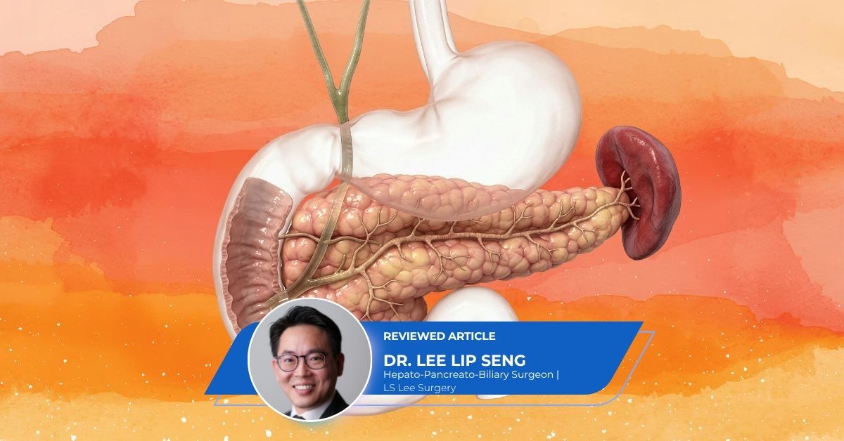
The detection of pancreatic cysts has increased in recent years, largely due to the increased accessibility of high-resolution abdominal imaging like Computed Tomography (CT) and Magnetic Resonance Imaging (MRI) in Singapore. While the word “cyst” can be alarming when associated with the pancreas, many of these fluid-filled sacs are benign. However, certain types of pancreatic cysts carry a risk of becoming cancerous, making it essential to distinguish between those that are harmless and those that require surgical intervention.
Get An Appointment With A Pancreas Surgeon
Contact UsBack To: Pancreas Cancer Main Page
What Is The Nature Of Pancreatic Cysts?
A pancreatic cyst is a pocket of fluid located on or within the pancreas. Unlike a solid tumour, which is a mass of tissue, a cyst is filled with either digestive enzymes or mucus. In clinical practice, these are broadly categorised into two groups: inflammatory pseudocysts and neoplastic cysts. Neoplastic cysts are further divided into those that are benign (non-cancerous) and those that are mucinous, which have the potential to develop into pancreatic cancer.
Serous Cystadenomas: Generally Benign
Serous Cystadenomas (SCAs) are a common type of neoplastic cyst. They are typically composed of many tiny, thin-walled sacs, often described as having a “honeycomb” appearance on imaging. These cysts are almost always benign and rarely, if ever, turn into cancer.
Most SCAs do not cause symptoms and are managed through periodic monitoring. Surgery is generally only recommended if the cyst grows large enough to compress surrounding organs, such as the stomach or bile duct, causing pain or digestive issues.
Mucinous Cystic Neoplasms: The Need For Caution
Mucinous Cystic Neoplasms (MCNs) are cysts filled with a thick fluid called mucin. These are most commonly found in the body or tail of the pancreas and occur predominantly in women. Unlike serous cysts, MCNs are considered “premalignant.” This means that while they may be benign at the time of discovery, they have a high potential to transform into invasive pancreatic cancer over time.
Because of this risk, Hepatopancreatobiliary (HPB) surgeons typically recommend the surgical removal of MCNs, especially in patients who are fit for surgery, to prevent future malignancy.
Intraductal Papillary Mucinous Neoplasms: Complex Management
Intraductal Papillary Mucinous Neoplasms (IPMNs) are another type of mucinous cyst that grow within the pancreatic ducts. They are categorised based on their location:
- Main-Duct IPMN: These involve the main pancreatic duct and carry a high risk of malignancy. Surgical resection is usually advised.
- Branch-Duct IPMN: These develop in the side branches of the duct system. They are often smaller and have a lower risk of turning into cancer compared to main-duct versions.
- Mixed-Duct IPMN: These involve both the main duct and the branches.
The management of IPMNs is highly personalised. Surgeons look for “worrying features,” such as a cyst size greater than three centimetres, a thickened cyst wall, or a dilated main pancreatic duct, to determine if surgery is necessary.
Pancreatic Pseudocysts: An Inflammatory Response
A pancreatic pseudocyst is not a true cyst because it is not lined by the type of cells found in neoplastic cysts. Instead, it is a collection of fluid and debris that forms as a complication of pancreatitis (inflammation of the pancreas).
Most pseudocysts resolve on their own with supportive care. However, if a pseudocyst becomes infected, ruptures, or grows large enough to cause a blockage, a surgeon may need to drain it using endoscopic or laparoscopic techniques.
How Are Dangerous Features In Pancreatic Cysts Identified?
When evaluating Pancreatic Cysts, specialists in Singapore use specific “red flag” criteria to identify which cysts are dangerous. These features include:
- Cyst Size: Cysts larger than three centimetres generally require closer scrutiny.
- Solid Components: The presence of a solid mass or “nodule” within the cyst significantly increases the suspicion of cancer.
- Ductal Dilation: A main pancreatic duct that is wider than five to ten millimetres often indicates a higher risk.
- Symptom Presentation: Cysts that cause jaundice (yellowing of the eyes and skin) or new-onset diabetes are treated with a higher level of urgency.
The Role Of Advanced Diagnostics
To accurately differentiate between these cysts, an HPB surgeon may recommend an Endoscopic Ultrasound (EUS). During this procedure, a fine needle can be used to sample the fluid inside the cyst (Fine Needle Aspiration). Analysing the fluid for tumour markers (such as CEA) and amylase levels helps the medical team provide a definitive diagnosis.
Consult A Pancreas Specialist
If the presence of pancreatic cysts has been detected, it is important to remain calm but proactive. Most cysts can be managed safely through regular surveillance. Consult a surgeon specialising in the pancreas to receive a formal assessment and a tailored management plan.
Contact UsBack To: Pancreas Cancer Main Page

This medical content has been reviewed by Dr Lee Lip Seng, a highly regarded surgeon who specialises in advanced minimally invasive robotic and laparoscopic techniques for the treatment of complex liver, pancreatic, and gallbladder conditions. Patients frequently seek his care due to his exceptional clinical expertise and a steadfast commitment to delivering compassionate, patient-centred results.
All content on this site is informative only and is not intended to be a substitute for professional medical advice, diagnosis, or treatment, and should never be relied upon for specific medical advice. If you are experiencing a medical emergency, call 995 immediately or go to the nearest Accident & Emergency department.
health365.sg is a member of Group 365Asia.